































Philips Verida Brings AI-Powered Spectral CT to Cancer Imaging
Philips Verida uses AI-powered spectral CT reconstruction to deliver images faster, supporting oncology, treatment planning and low-dose lung cancer screening.
Cancer imaging often comes down to two things that do not always sit comfortably together: detail and speed.
Radiologists need images clear enough to distinguish subtle abnormalities, while hospitals also need scanners that can move patients through increasingly busy imaging departments without adding unnecessary delays.
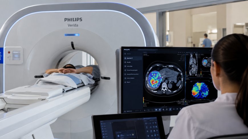
Philips is addressing both with Verida, its latest spectral CT system.
The company received U.S. FDA 510(k) clearance for the Spectral CT Verida family in March 2026 and formally announced it in April. The system combines Philips’ detector-based spectral CT technology with AI-powered image reconstruction. It is intended for a range of diagnostic applications, including oncology and low-dose CT lung cancer screening within established screening protocols.
Read Philips’ official Verida FDA clearance announcement.
The interesting part is not that an AI system is independently looking at a scan and declaring whether a patient has cancer.
It is what happens before the radiologist begins interpreting the images.
AI Is Built Into the Imaging Chain
Verida uses Philips’ Spectral Precise Image technology, a deep-learning reconstruction system designed to reduce image noise while preserving anatomical detail.
That reconstruction step matters.
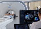
CT scanners collect raw X-ray measurements that have to be turned into images before a radiologist can interpret them. Better reconstruction can help produce clearer images while reducing artefacts and potentially allowing useful information to be obtained at lower radiation doses.
Philips has built AI into that process rather than adding it as a separate application after the examination.
The company’s third-generation dual-layer detector captures high- and low-energy X-ray information during the same acquisition. That allows the system to produce conventional CT images and spectral information from one examination.
See the Philips Verida product page.
For oncology, spectral imaging can be useful because different tissues and materials interact with X-rays differently. The additional information can help clinicians characterise findings that may be harder to distinguish on conventional CT alone.
That does not make the scanner an automated cancer diagnosis system. A radiologist still has to interpret what the images mean.
The Speed Claim Is About Reconstruction
One of VVerida’s most notable specifications is how quickly it processes images after an examination.
Philips says the system can reconstruct 145 images per second, allowing an entire exam to appear automatically in less than 30 seconds.
The company describes that as roughly twice as fast as its previous generation and says the system can support as many as 270 examinations per day under its stated workflow assumptions.
Those numbers need to be understood correctly.
They do not mean every patient is physically scanned for cancer in 30 seconds.
The less-than-30-second figure refers to the time for the complete examination images to become available after reconstruction.
That distinction matters, especially in healthcare technology where marketing language around AI and speed can easily be misunderstood.
Still, faster reconstruction has practical value.
If images are available sooner, radiologists can begin reviewing them sooner, while high-volume departments may spend less time waiting for image processing between examinations.
Verida Has a Specific Lung Cancer Screening Use
The cancer angle is not limited to general oncology imaging.
Philips states that Verida is intended for low-dose CT lung cancer screening when used within established screening protocols.
Low-dose CT is already an important screening tool for people at elevated risk of lung cancer.
AI-powered reconstruction is particularly relevant here because lung screening has to balance image quality with radiation exposure.
A reconstruction system that can suppress image noise while maintaining clinically useful detail may help support that balance.
Again, Verida should not be confused with AI software that automatically finds lung cancer.
Its role is primarily to acquire and reconstruct CT images that clinicians then evaluate.
The FDA record confirms that the Spectral CT Verida Family received 510(k) clearance on March 27, 2026, as a Class II computed tomography X-ray system.
View the FDA 510(k) record for Philips Verida
Spectral CT Can Provide More Than a Conventional Image
Traditional CT produces images largely based on differences in how tissues absorb X-rays.
Spectral CT goes further by collecting information at different X-ray energy levels.
That can help radiologists distinguish materials and tissues that may look similar on a conventional scan. Verida’s detector collects high- and low-energy information during a single acquisition, so spectral data is available without a separate examination.
Philips describes this as “always-on” spectral imaging.
The practical benefit is that a radiologist may have access to additional information even when a spectral analysis was not specifically requested before the patient was scanned.
That can be useful in oncology because radiologists sometimes need more information about a lesion after the patient has already left the scanner.
Avoiding an additional scan saves time and reduces unnecessary repeat imaging when the existing spectral data answers the question.
The Scanner Is Also Built for Cancer Treatment Planning
Cancer imaging is not only about finding a tumour.
CT is also used extensively to plan radiation therapy.
Verida is cleared for oncology applications, including treatment preparation and radiation-therapy planning.
The system supports extended field-of-view imaging and respiratory-correlated 4D CT for treatment planning and simulation.
That is important for tumours affected by breathing motion, particularly in areas such as the chest.
Radiation-oncology teams need to understand not only where a tumour is located, but how its position can change as the patient breathes.
A faster imaging and reconstruction workflow can therefore matter at several stages of cancer care, from screening and diagnosis to treatment planning.
Philips Is Putting AI Into More of Its Medical Equipment
Verida is not an isolated AI project for Philips.
The company has been integrating AI into ultrasound, MRI, CT and other imaging systems.
TechAmerica.ai recently covered the company’s Alturion ultrasound platform and its AI-assisted clinical workflows.
The pattern is similar.
Instead of building a separate AI application that clinicians have to open after using the medical device, Philips is putting automation directly into the equipment and imaging workflow.
With Alturion, that includes automated ultrasound measurements.
With Verida, the AI is working deeper in the CT imaging chain, particularly during image reconstruction.
That approach may ultimately be more useful in hospitals than adding another standalone AI dashboard for clinicians to manage.
AI Does Not Replace the Radiologist
It is tempting to describe an AI-powered CT scanner as a machine that “detects cancer faster.”
That would overstate what Verida actually does.
The system is a CT scanner, not an autonomous cancer-detection model.
AI helps reconstruct the images, suppress noise and support an efficient imaging workflow. Spectral CT provides additional information that may help clinicians characterise tissue and evaluate subtle findings.
But the radiologist remains responsible for interpreting the examination.
That difference is important.
Some healthcare AI products are specifically designed to flag suspicious lesions or calculate cancer risk. Verida’s primary AI function is earlier in the process: helping turn acquired CT data into usable images quickly and with high image quality.
Its value in cancer care therefore comes from giving clinicians better imaging information sooner, rather than making the diagnosis for them.
Why Faster CT Matters
Cancer imaging departments are dealing with growing examination volumes while many healthcare systems face staffing pressure.
Speed alone is not enough. A fast scan is of little value if image quality is poor.
That is why combining reconstruction speed and image quality matters more than either metric in isolation.
Philips says Verida’s AI-based reconstruction can reduce noise while preserving fine detail. Its spectral detector can also produce conventional and spectral information from the same acquisition.
If those capabilities reduce repeat imaging or shorten the time before a radiologist can confidently interpret an examination, the benefit is operational as well as clinical.
The company says its existing detector-based spectral CT technology is already used across hundreds of installed systems globally, giving Verida a foundation in technology that is already part of routine clinical imaging rather than a purely experimental platform.
Cancer Imaging Is Becoming More Software-Driven
CT scanners have traditionally been judged by hardware: detector performance, scan speed, X-ray output and spatial resolution.
Those factors still matter.
But software is becoming just as important.
The quality of the reconstruction algorithm can affect what a radiologist sees. Automated workflows can determine how quickly an exam is processed. Spectral processing can extract additional information from the same underlying scan.
That makes Verida an interesting example of where medical imaging is heading.
The machine is still fundamentally a CT scanner.
What has changed is how much intelligence sits between the raw data the detector collects and the image that appears on the radiologist’s screen.
For cancer care, that may be where AI proves most useful in the near term: not replacing the physician who reads the scan, but helping deliver better information to that physician faster.
What's Your Reaction?
 Like
0
Like
0
 Dislike
0
Dislike
0
 Love
0
Love
0
 Funny
0
Funny
0
 Angry
0
Angry
0
 Sad
0
Sad
0
 Wow
0
Wow
0